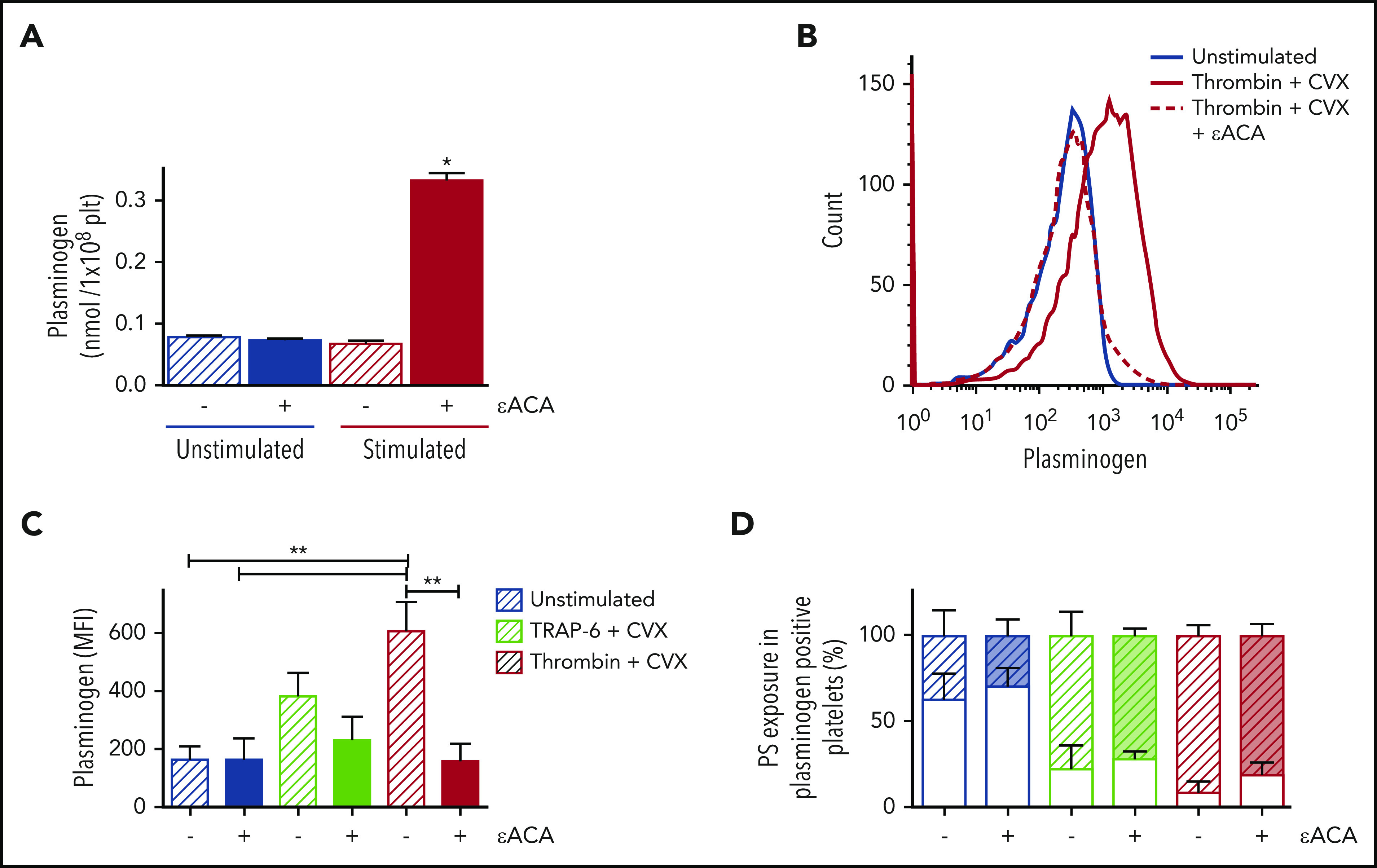
Figure 1.

Plasminogen is released from activated platelets and retained on the stimulated membrane. (A) Isolated human platelets were stimulated with collagen (20 µg/mL) and thrombin (100 nM) in the absence (hatched bars) or presence of εACA (200 mM, solid bars) for 30 minutes. Plasminogen was detected in the supernatant by ELISA. (B-D) Platelets (2 × 10⁸ platelets/mL) were stimulated ± CVX (100 ng/mL) with TRAP-6 (15 μM) or thrombin (100 nM) ± εACA. Annexin V-AF488 and anti-plasminogen antibody-DL633 were included to detected PS exposure and platelet-derived plasminogen, respectively. (B) Representative flow cytometry curves. Data are presented as mean ± SEM for (C) MFI for platelet-derived plasminogen exposure and (D) PS exposure in plasminogen-positive platelets. PS-negative platelets are indicated by hatched bars and PS positive by open bars. Color coding as in panel C. *P < .05, **P < .01; n ≥ 3.
