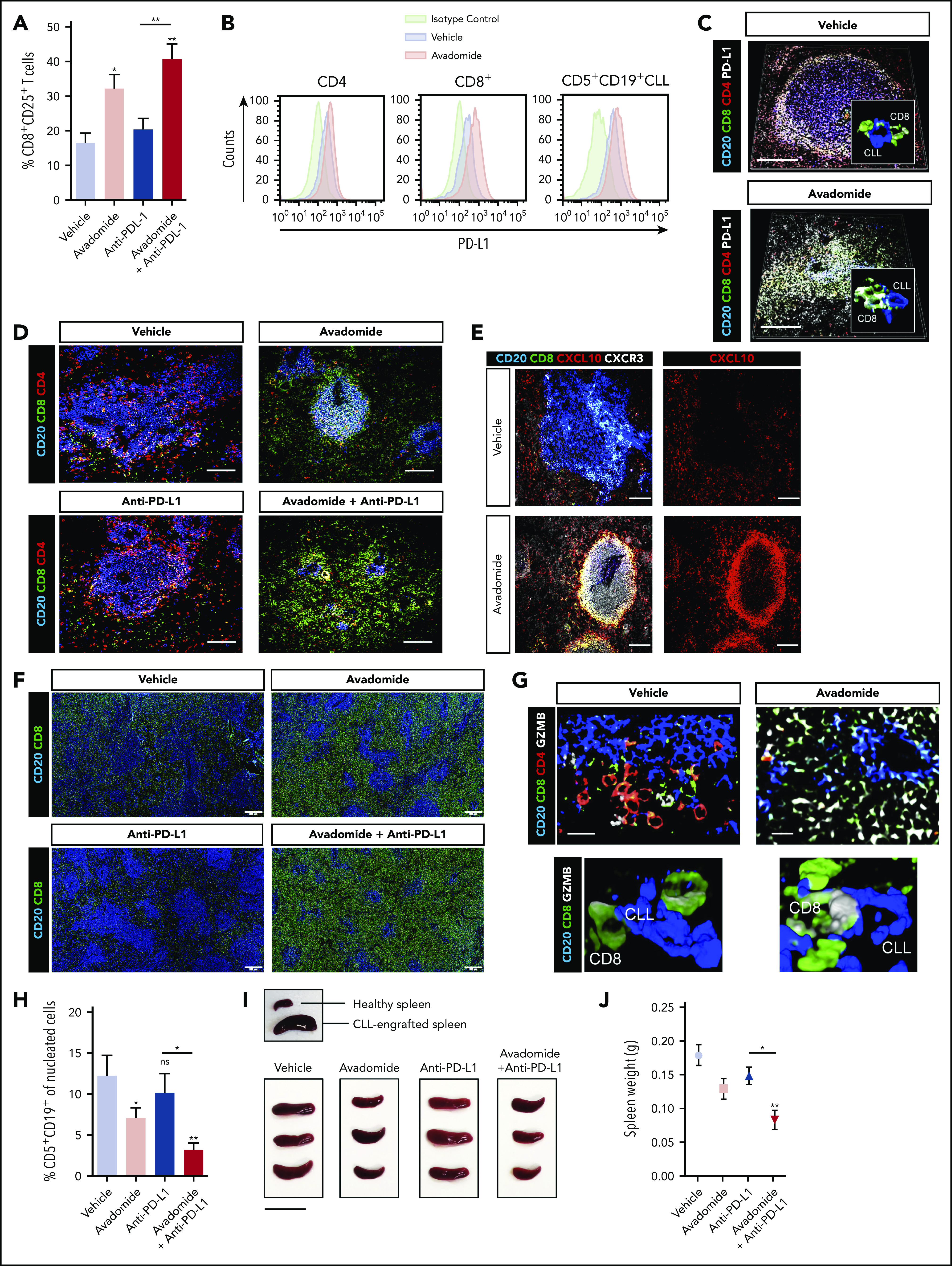
Figure 6.

Therapeutic avadomide converts CD8+ T cell excluded (noninflamed) patient-derived xenografts into CD8+ T cell-inflamed tumors that respond to anti-PD-L1 combination therapy. (A) Flow cytometric percentage of patient CD8+ CD25+ T cells harvested from CLL patient-derived xenograft splenic TMEs following drug treatments (n = 6 patient samples, 3-4 mice per patient sample treatment group). (B) Representative flow cytometric histograms of PD-L1 expression on patient CD4+, CD8+ T cells, and CD5+ CD19+ CLL cells harvested from the splenic TME comparing vehicle (blue) and avadomide (red) treated mice. Representative multispectral immunofluorescence images of splenic TME tissue (n = 6 patient samples) from treated mice (C) for human CD20 (blue), CD8+ (green), CD4+ (red) patient T cells and PD-L1 (white); (D) for human CD20 (blue), CD8+ (green) and CD4+ (red) patient T cells; (E) for human CD20 (blue), CD8+ (green), CXCL10 (red), and CXCR3 (white); (G) for human CD20 (blue), CD8+ (green), CD4+ (red) patient T cells, and granzyme B (GZMB) (white). Original magnification, ×20 medial optical section images (scale bar = 100 μm for panels C-E and 20 μm for panel G) and 3D volume rendered confocal images of intercellular PD-L1+ (C) or GZMB+ (G) CD8+ T cell interactions (white/green) with CLL cells (blue) with treatments (cropped, ×20 images). (F) Representative large images acquired by an Olympus BX61VS fluorescence slide scanner (original magnification, ×4, scale bar = 200 μm) of splenic TME tissue (n = 6 patient samples) from treated mice for human CD20 (blue) and CD8+ (green) patient T cells. (H) CLL tumor burden in splenic TMEs. Flow cytometric percentage of human CD5+ CD19+ CLL cells of tissue splenocytes (total nucleated cells, human and murine) (n = 6 patient samples, 3-4 mice per patient sample treatment group) analyzed from splenic TMEs following drug treatments. (I) Representative pictures of patient-derived xenograft splenic TME tissues. An established tumor (CLL PBMC engrafted spleen) in comparison with a nondiseased healthy murine spleen is shown (top). Xenograft splenic TME tissues are shown following different treatments (scale bar = 2 cm). (J) Weight of xenograft (n = 3 patient samples) spleen TME tissues following drug treatments. *P < .05; **P < .01; ns, not significant using a repeated measures 1-way ANOVA with Tukey's multiple comparisons test (A,H,J). Data presented as mean ± SEM.
