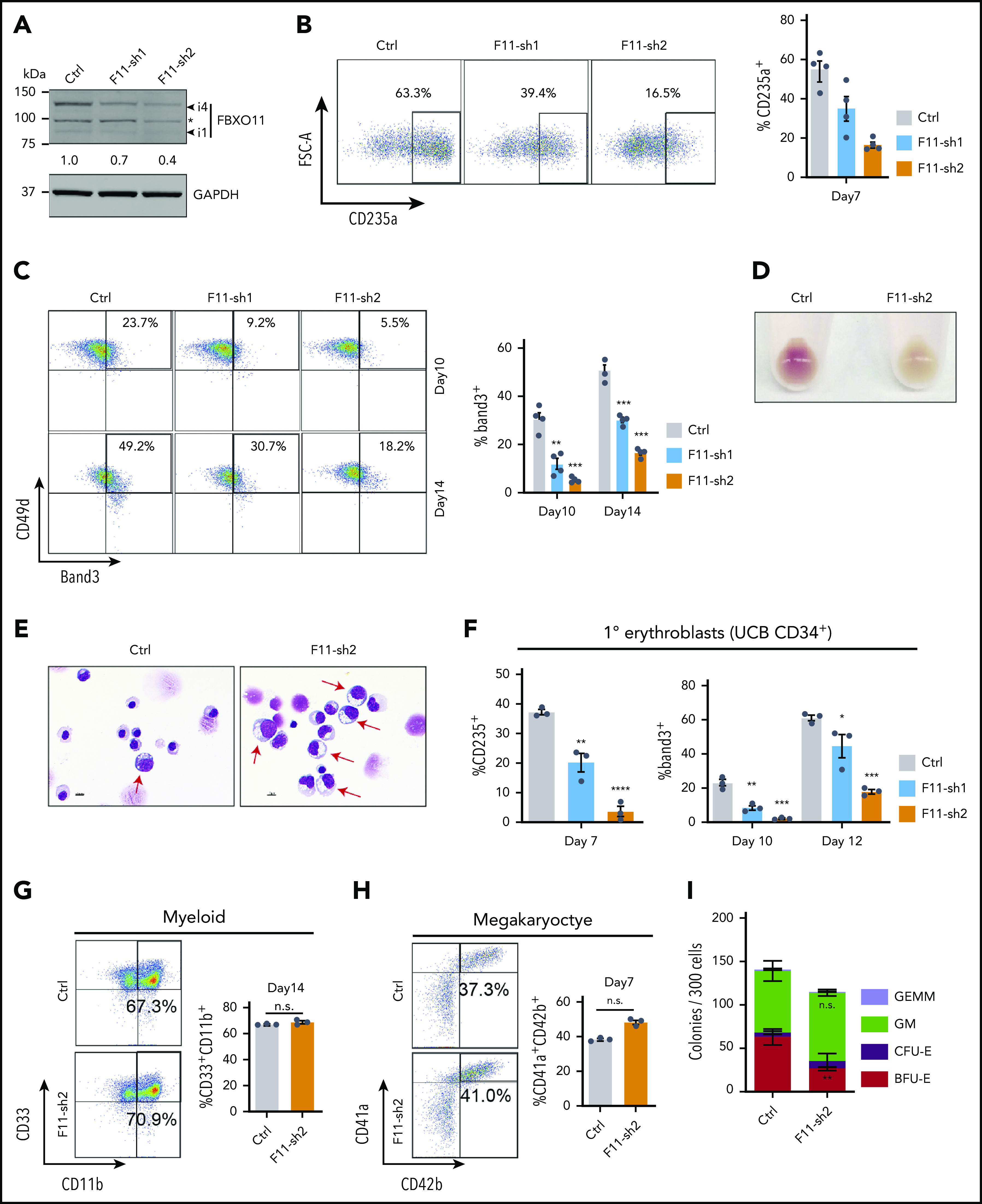
Figure 2.

FBXO11 regulates human erythropoiesis. Peripheral blood–mobilized human CD34+ cells (A-E,G-I) were expanded for 2 days and then transduced with lentiviral vectors encoding green fluorescent protein (GFP) and FBXO11 (F11-sh) or control (Ctrl) shRNAs. At day 4, FACS-purified GFP+ cells were cultured under conditions supporting erythroid (A-F), myeloid (G), megakaryocyte (H), or multilineage (I) differentiation. (A) Western blot showing FBXO11 protein expression at culture day 10. The asterisk denotes a nonspecific band. The number below shows quantification of the major isoform of FBXO11 (i4) normalized to GAPDH protein for each condition. (B) Flow cytometry plots showing the expression of the erythroid marker CD235a at culture day 7. The graph shows the mean ± SEM from 4 biological replicate experiments. (C) Flow cytometry plots showing the expression of Band3 and CD49d at culture days 10 and 14. The graph shows the mean ± SEM from 4 biological replicate experiments. (D) Cell pellets at day 10. (E) May-Grünwald-Giemsa–stained erythroblasts at day 14. Red arrows denote immature erythroblasts. Scale bar, 10 μM. (F) Umbilical cord blood (UCB) CD34+ cells expressing FBXO11 (F11-sh) or Ctrl shRNAs were cultured in erythroid cytokines. Graphs show the fraction of cells expressing CD235a or Band3 (mean ± SEM) at the indicated time points in 3 biological replicate experiments. (G) Peripheral blood CD34+ cells expressing FBXO11 (F11-sh) or Ctrl shRNAs were cultured with myeloid cytokines. Flow cytometry plots show the expression of CD11b and CD33 at culture day 14. The graph shows the mean ± SEM from 3 biological replicate experiments. (H) Peripheral blood CD34+ cells expressing FBXO11 (F11-sh) or Ctrl shRNAs were cultured with megakaryocytic cytokines. Flow cytometry plots show the expression of CD41a and CD42b at day 7. The graph shows the mean ± SEM from 3 biological replicate experiments. (I) A total of 300 peripheral blood CD34+ cells expressing Ctrl or F11-sh2 shRNA were suspended in 1 mL of methylcellulose containing multilineage cytokines. Hematopoietic colonies were enumerated after 14 days. The graph shows the mean ± SEM from 3 replicate experiments. BFU-E, burst-forming unit-erythroid; CFE-E, colony-forming unit-erythroid; GEMM, granulocyte, erythroid, macrophage, megakaryocyte; GM, granulocyte macrophage. *P < .05; **P < .01; ***P < .001; ****P < .0001 relative to shRNA luciferase (shLuc) (unpaired Student t test).
