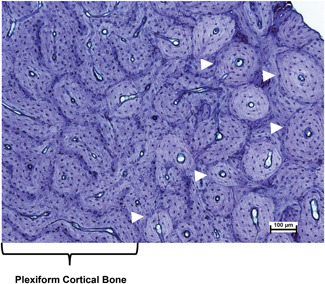
Figure 2.

Photomicrograph of an undecalcified section of ovine endosteal cortical bone from the radius. Plexiform cortical bone is on the left. Active remodeling is indicated by the presence of secondary osteons (white arrow heads) on the right. Toluidine blue stain. Scale bar = 100 µm. (Photo courtesy of Dr. Clifford Les) [Color figure can be viewed at wileyonlinelibrary.com]
