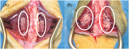
Figure 4.

Demineralized bone matrix (A, white circles) and corticocancellous bone (B, white circles) during implantation in a rat model of lumbar spinal fusion. The lumbar spine is evident between the circles in each image [Color figure can be viewed at wileyonlinelibrary.com]
