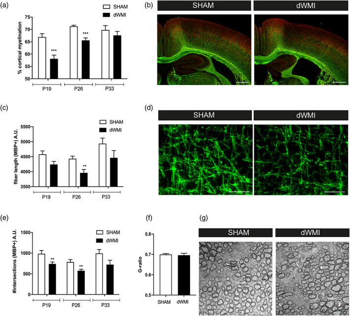
FIGURE 1.

The combination of postnatal hypoxia/ischemia and systemic inflammation at P5 (dWMI model) causes a delay in myelination in neonatal mice. (a) Mice exposed to the double‐hit model displayed a transient reduction in cortical myelination (P19 SHAM n = 11 dWMI n = 11, P26 n = 7 in both groups, P33 SHAM n = 4 dWMI n = 5). (b) Representative fluorescent images (×2.5) of the ipsilateral cortex of a sham‐operated control mouse (left) and dWMI mouse (right) stained for axonal marker NF200 (red) and myelin marker MBP (green). Scale bars: 500 μm. (c, e) Microstructural changes in MBP+ fibers following dWMI induction were observed at P19 and P26, assessed by measuring fiber length (c) and number of intersections (e) (P19 SHAM n = 11 dWMI n = 11, P26 n = 8 in both groups, P33 SHAM n = 4 dWMI n = 5). (d) Representative fluorescent images (×40) of MBP+ axons in the ipsilateral cortex of a sham‐operated control mouse (left) and dWMI mouse (right). Scale bars: 100 μm (f) G‐ratio analyses (axonal diameter divided by the fiber diameter including the myelin sheath) reveal no changes in myelin enwrapment at P33 in SHAM (n = 3) and dWMI (n = 4) animals. (g) Representative electron microscopy images of the caudal corpus callosum in SHAM and dWMI animals at P33. Scale bars: 2 μm. **p < .01; ***p < .001 sham‐operated control versus dWMI animals at the specified timepoint [Color figure can be viewed at wileyonlinelibrary.com]
