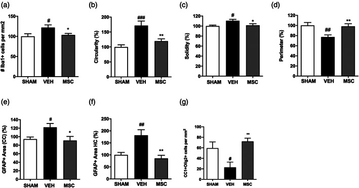
FIGURE 8.

Intranasal MSC treatment dampens the neuro‐inflammatory response and restores OL maturation following dWMI (a) Quantification of microglia density in the corpus callosum revealed a reduction of Iba1 + cells after MSC treatment compared with vehicle treatment (SHAM n = 9, VEH n = 10, MSC n = 14). (b–d) Microglia morphology analyses assessed by cell circularity (b), solidity (c), and perimeter (d), showed a less pro‐inflammatory phenotype following MSC treatment compared with vehicle treatment (SHAM n = 9, VEH n = 11, MSC n = 12, normalized to control values). (e,f) A reduction in GFAP+ area in the corpus callosum (e) and hippocampus (f) was observed following intranasal MSC treatment compared with vehicle‐treatment (SHAM n = 8, VEH n = 8, MSC n = 8, normalized to control values). G. Intranasal administration of 0.5 × 10⁶ MSCs restored CC1+/Olig2+ cells numbers up to sham‐control levels in dWMI animals, indicating a boost in OL lineage maturation (SHAM n = 6, VEH n = 6, MSC n = 13). #p < .05; ##p < .01; ###p < .001 vehicle‐treated dWMI animals versus sham‐controls; *p < .05; **p < .01 MSC‐treated dWMI versus vehicle‐treated dWMI animals
