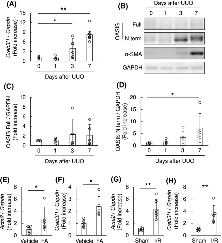
FIGURE 2.

Expression of OASIS was upregulated in murine fibrotic kidneys. A, Total RNA was prepared from the murine kidneys after UUO. The expression of OASIS/Creb3l1 transcript was analyzed at indicated time points after UUO by quantitative RT‐PCR. The expression of the transcript was normalized to that of Gapdh. Data are shown as mean ± SD (n = 6), ** P < .01 vs. Day 0 by one‐way ANOVA followed by Dunnett test. B, The kidney lysates from mice at 0, 1, 3 and 7 days after UUO were immunoblotted with anti‐OASIS, anti‐α‐SMA and anti‐GAPDH antibodies. Representative images are shown. C and D, Quantitative analysis for protein expression levels of OASIS in kidneys after UUO. Data are shown as mean ± SD (n = 5), * P < .05 vs. Day 0 by one‐way ANOVA followed by Dunnett test. E‐H, The expression of Acta2 (E, G) and OASIS/Creb3l1 (F, H) transcripts was estimated at 2 weeks after folic acid treatment (E, F; n = 5) or at 3 weeks after ischemia and reperfusion (I/R) injury (G, H; n = 6). The expression of the transcripts was normalized to that of Gapdh. Data are shown as mean ± SD, * P < .05, ** P < .01 vs. vehicle or sham by student's t test
