
Fig 2.
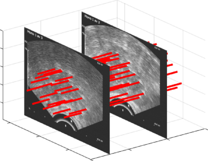
Ultrasound images encompassing the complete prostate gland with margins, acquired after a sweep of the transrectal ultrasound, yielding a three‐dimensional volume including 20 needles (highlighted with red lines). [Color figure can be viewed at wileyonlinelibrary.com]