
Figure 3.
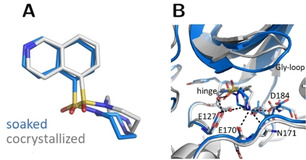
Crystal structures of fasudil (1) in complex with PKA from a soaked (blue, PDB‐code: 6YNA) and cocrystallized crystal (gray, PDB‐code: 5LCP). A: Ligand superposition indicates a slight rotation of the ligand between both structures (RMSD ligand: 1.0 Å, RMSD of the Cα atoms of the hinge region: 0.5 Å). B: Active site superposition. Dotted lines indicate hydrogen bonds. The Gly‐rich loop is positioned with a more open geometry in the cocrystal structure and the latter structure shows more hydrogen bonds between the ligand and PKA.