
Figure 6.
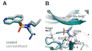
Crystal structures of open chain fasudil‐derivative 4 in complex with PKA from a soaked (teal, PDB‐code: 6YQJ) and cocrystallized structure (gray, PDB‐code: 5LCR). (A) Ligand superposition indicates a slight shift of the entire ligand as well as an altered position of the aminoethyl moiety in the two structures (RMSD ligand: 0.9 Å, RMSD of the Cα atoms of the hinge region: 0.4 Å). (B) Active site superposition. Dotted lines indicate hydrogen bonds. The position of the Gly‐rich loop is not visible in the soaked crystal structure. The hydrogen‐bonding pattern differs between the structures obtained by the two protocols. In the cocrystal structure hydrogen bonds are formed to the hinge, the side chains of Asp184 and Asn171 and the backbone of Glu170. In the soaked structure only the hinge and the backbone of Glu170 are involved in H‐bonding and an additional hydrogen bond is established to the side chain of Glu127 in the soaked structure.