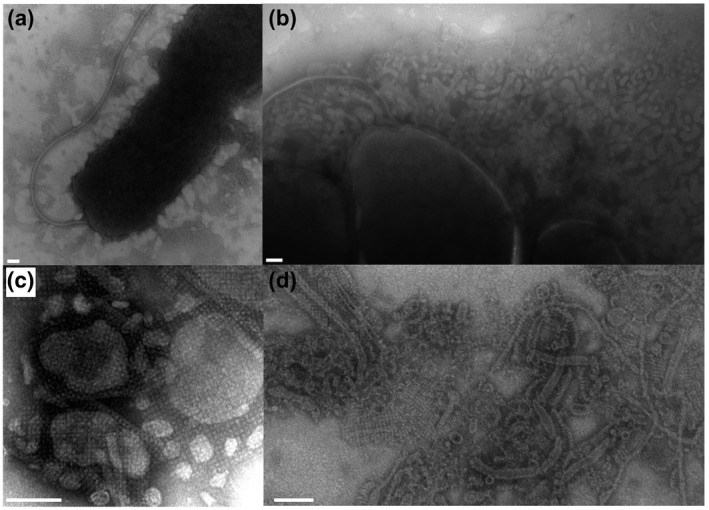
FIGURE 5.

S‐layer is present in the P. tunicata extracellular environment and biofilm matrix. (a) A TEM micrograph showing a P. tunicata cell as well as “shed” extracellular material layer that is nearby but distinct from the cell body. (b) Three adjacent cells within a microcolony with a substantial amount of secreted outer membrane vesicles (OMVs) and a sheathed flagellum. (c) TEM micrograph of an extracellular protein fraction showing S‐layer associated with outer membrane vesicles of various forms. (d) TEM image taken from a WT pellicle biofilm, which shows S‐layer material in association with outer membrane vesicles, and fibrous and tubular structures. (e.g., see high‐magnification image shown in d) representing possible pili, flagella, prosthecae, or flagellar sheaths. Scale bars = 100 nm
