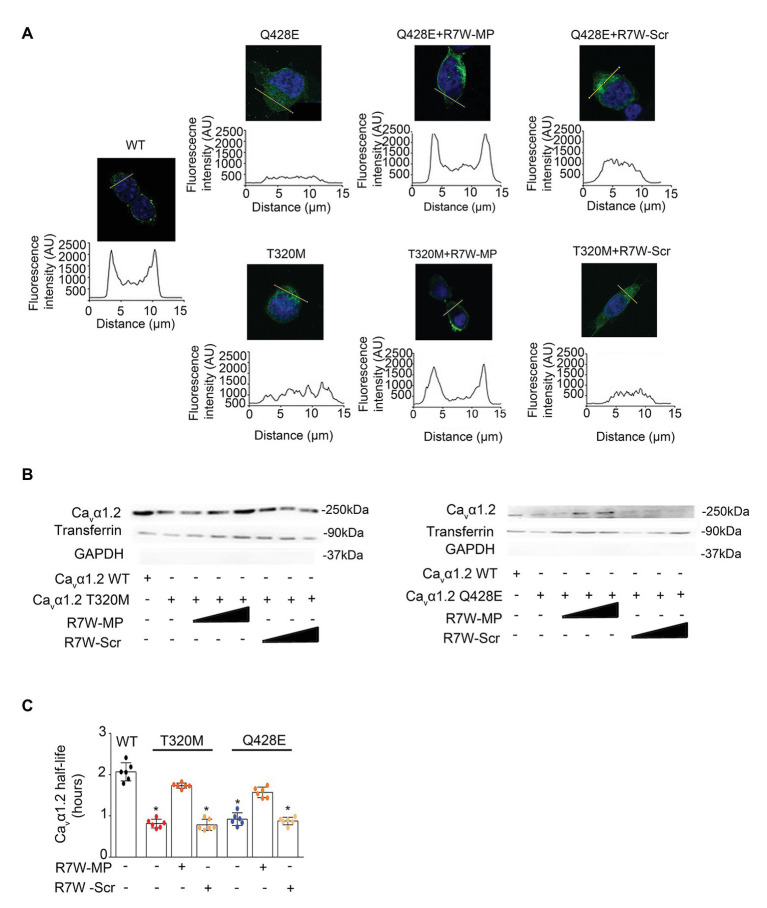
Figure 4.
R7W-MP corrects LTCC density at the plasma membrane in Cavα1.2 mutant-transfected cells. (A) Subcellular localization of Cavα1.2 (top) and line scan analyses (bottom) for each condition as indicated. AU, arbitrary units. Scale bars (in white), 10 μm (n = 50). Representative experiments are shown. Cells were treated with increasing doses (0.12, 1.3, and 10.2 μM) of R7W-MP or R7W-Scr. (B) Cell surface biotinylation assay followed by followed by Western blot analysis (a representative image is shown) on transfected cells treated as indicated (n = 3). (C) Cavα1.2 half-life as measured in a NanoLuc luciferase assay. HEK293 cells were transfected with Cavα1.2-NanoLuc (WT, T320M, and Q428E mutant) and treated with 1.3 μM R7W-MP or R7W-Scr as indicated (n = 6). * p < 0.05 (Mann-Whitney test).

