FIG. 5.

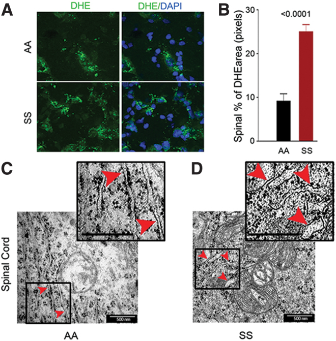
Spinal cords of sickle mice have increased ROS and ER stress. (A) Representative images of L4 and L5 spinal sections from HbSS-BERK and HbAA-BERK mice stained for ROS formation (DHE, green) and nuclei (DAPI, blue). Magnification 600 × ; bar = 10 μm. Each image is representative of images from 8 mice. (B) ROS expression as percentage of DHE-positive pixels was obtained from the right and left dorsal horn of each section (n = 8 per group). (C, D) Representative TEM images of ER (red arrowheads) in the spinal cords (magnification 60,000 × , bar = 500 nm) from the L4–L5 region. HbSS-BERK sickle mice exhibit pronounced swelling of ER cisternae (D) as compared with the normal, narrow ER cisternae in HbAA-BERK control normal mice (C). Insets (demarcated with boxes) show enhanced details of cisternae swelling. Each image is representative of images from three mice. DAPI, 4′,6-diamidino-2-phenylindole; DHE, dihydroethidium; TEM, transmission electron microscopy. Color images are available online.
