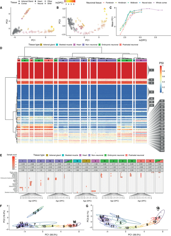
Fig. 3.
Microexon inclusion through mouse embryonic development. a, b Dimensionality reduction using probabilistic principal component analysis of microexon PSI values across mouse embryonic and postnatal samples reveals correlation with developmental time for PC1, while PC3 is correlated with the developmental time of the postnatal brain samples. Dot shapes denote different sample types, and their colour indicate their developmental stage, here expressed as log days postconception (DPC). c PC1 correspondence with embryonic developmental time. d Heatmap showing the microexon inclusion patterns across analysed RNA-seq samples, where rows correspond to microexons and columns to RNA-seq samples. Blue to red colour scale represents PSI values. RNA-seq samples were clustered in 16 different clusters. Microexons were clustered in 24 different clusters, and these were named according to their further classification as neuronal (N), muscular (M), neuromuscular (NM), weak-neuronal (WN) and non-neuronal (NN). e Tissue type and developmental stage composition from sample clusters containing neuronal samples or samples associated with high microexon inclusion. f, g Projection of the sample clusters across the estimated principal components

