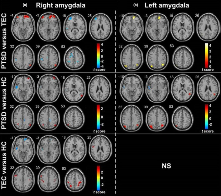
Figure 3.

Group differences in whole amygdala resting‐state functional connectivity maps. (a) and (b) represent the group differences in right and left whole amygdala functional connectivity, respectively (p < .001, cluster >10, uncorrected). Warm color represents the positive functional connectivity; cold color represents the negative functional connectivity.Note: HC, healthy control group; PTSD, post‐traumatic stress disorder group; TEC, trauma‐exposed control group
