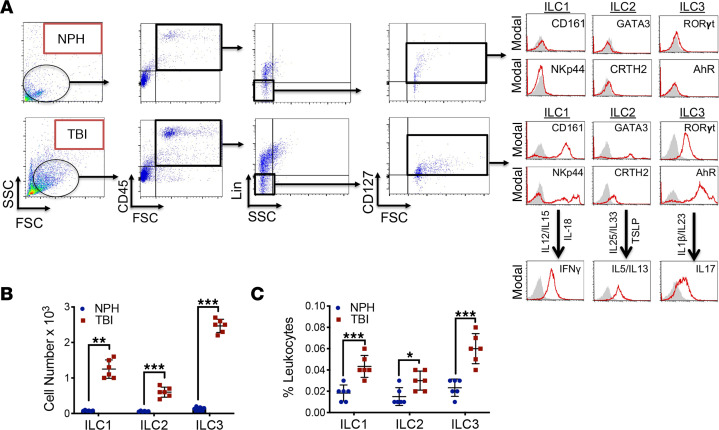
Figure 2. Increased presence of ILC1 and ILC3 within human CSF after TBI.
(A) CSF was collected from consecutive, adult nontraumatic control (normal pressure hydrocephalus; NPH) or severe TBI patients. Human ILCs were sorted using forward scatter (FSC)/side scatter (SSC) and identified as CD45+, lineage-negative (Lin−), CD127+ lymphoid cells. Selected populations were analyzed for ILC subtype as follows: ILC1, Lin−CD127+CD161+NKp44+; ILC2, Lin−CD127+GATA3+CRTH2+; and ILC3, Lin−CD127+AhR+RORγt+. ILC functionality was further assessed by cytokine production (ILC1, IFN-γ; ILC2, IL-5/IL-13; and ILC3, IL-17) after cytokine stimulation, as shown. Gray shaded areas indicate isotype controls. (B and C) Quantified data reveal low basal expression of ILC subtypes, with large increases in all ILC classes after TBI. Scatterplots, which are expressed as mean ± SD, depict ILC subtypes as total cell number (B) and % leukocytes (C). Data from individual patients (n = 6 NPH patients, n = 6 severe TBI patients) were compared within each ILC subtype using a 2-tailed Student’s t test (*P < 0.05, **P < 0.01, ***P < 0.001 versus sham).

