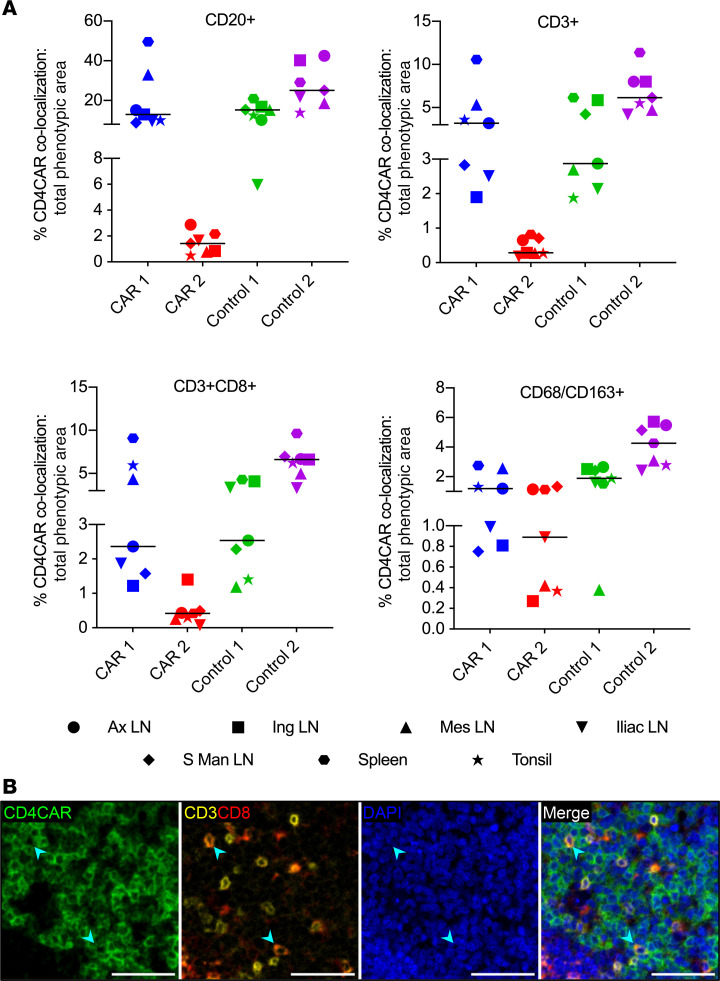
Figure 4. Multilineage engraftment of HSPC-derived CAR+ cells in lymphoid germinal centers.
(A) Percentages of total B cell (CD20+), T cell (CD3+), CTL (CD3+CD8+), and monocyte/macrophage (CD68/CD163+) immunophenotypic area that colocalized with CD4CAR immunoreactivity in GCs (n = 4 macaques; 6–7 lymphoid tissues per macaque). The charts show individual data points with medians. (B) Representative fluorescent mIHC photomicrographs of CD4CAR (green), CD3 (yellow), CD8 (red), and DAPI nuclear counterstain (blue), from CAR 1 mesenteric LN. Arrowheads indicate colocalization between CD4CAR and CD3/CD8 markers, showing the presence of CD4CAR+ CTLs within the germinal center. Scale bars: 50 μm.

