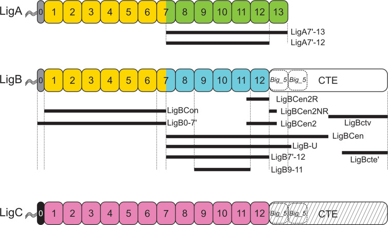
Figure 1.
Leptospiral Immunoglobulin-Like (Lig) Protein Immunoglobulin-like Domain Structure. Domain structures of the three Lig proteins are shown. LigA, LigB, and LigC contain a series of 13, 12, and 12 Big_2 domains, respectively. The first six Big_2 domains and part of the seventh domain of LigA and LigB have a high percentage of sequence identity as indicated by yellow domains. The sequences of the remaining Big_2 domains differ between LigA (green) and LigB (blue). The Big_2 domains of LigC are shown in pink. The Big_2 domains of LigB and LigC are followed by a C-terminal extension (CTE) predicted to harbor Big_5 domains (dashed rectangle). The “0” segment between the amino terminus of the mature proteins and Big_2 domain 1 is indicated with gray or black shading. All three Lig proteins are thought to have triacylated amino-terminal cysteines. Lig fragments are shown with black lines, and their amino acid coordinates are: LigA7’-13 (631–1224), LigBCon (47-630), LigBCen (631-1417), LigBCen2R (1014-1124), LigBCen2NR (1120-1165), LigBCen2 (1014-1165), LigBCen2NR (1014-1124), LigBCen2NR (1120-1165), LigB7’-12 (631-1124), LigB-U (631-1257), LigBctv (1418-1889), LigBcte’ (1630-1890). Lig fragment nomenclature may differ from that used in the original studies.

