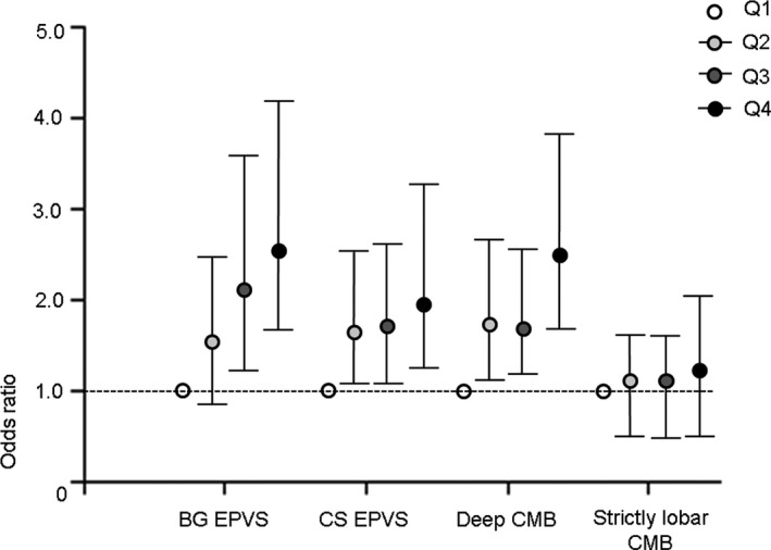
FIGURE 2.

Relationship between arterial stiffness and cerebral small vessel disease markers at different locations. When the patients were grouped according to quartiles of brachial‐ankle pulse wave velocity, enlarged perivascular spaces (EPVS) at the basal ganglia and centrum semiovale and deep CMB showed similar patterns of increase. BG EPVS (OR per baPWV quartile, Q2: 1.63, 95% CI 0.92–2.88; Q3: 2.11, 1.20–3.71; and Q4: 2.58, 1.45–4.60). CS EPVS (OR per baPWV quartile, Q2: 1.71, 1.07–2.73; Q3: 1.72, 1.06–2.78; and Q4: 2.06, 1.24–3.42). Deep CMB (OR per baPWV quartile, Q2: 1.74, 1.10–2.73; Q3: 1.70, 1.08–2.69; and Q4: 2.52, 1.62–3.94). Strictly lobar CMB (OR per baPWV quartile, Q2: 1.13, 0.69–1.85; Q3: 1.14, 0.68–1.93; and Q4: 1.30, 0.73–2.30). The cutoff values of mean brachial‐ankle pulse wave velocity for each quartile were 1,663, 1,985, and 2,355 cm/s. BG, basal ganglia; CMB, cerebral microbleed; CS, centrum semiovale; PVS, perivascular space
