Fig. 1.
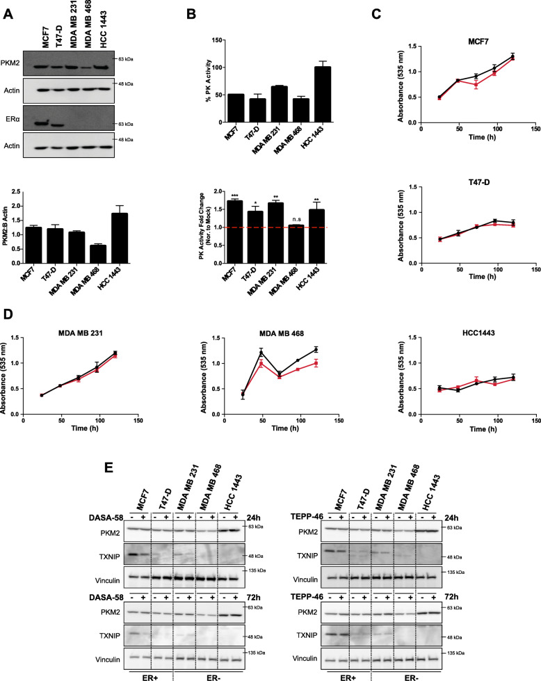
PKM2 pharmacological activation enhances pyruvate kinase activity in breast cancer cells without a clear effect on proliferation. a Representative western blot showing different PKM2 expression levels in five breast cancer cell lines (upper panel) with a densitometric analysis showing levels of PKM2 normalized to actin values (lower panel). b PK basal activity in BCa cells represented as percentage PK activity (PK activity in HCC1443 is considered 100%) measured in crude cell extracts (upper panel), PK activity levels in response to DASA-58 (15 μM), data shown as fold change in activity normalized to mock treatment, all tested cell lines except MDA MB 468 respond to DASA-58 treatment by increasing PK activity as measured in crude cell extracts (lower panel). c, d Total protein staining using SRB assay showing no significant change in cell survival in response to DASA-58 (15 μM; red line) in comparison to mock treatment (black line) in ER+ BCa cells (c) and ER − BCa cells (d) for up to 5 days under standard culture conditions. e Western blots showing the expression of PKM2 and TXNIP in response to either DASA-58 (15 μM) or TEPP-46 (30 μM) in short (24 h) and long (72 h) treatments. PKM2 activators do not change PKM2 levels in the tested cell lines but seemingly reduce TXNIP levels in cells expressing detectable TXNIP levels

