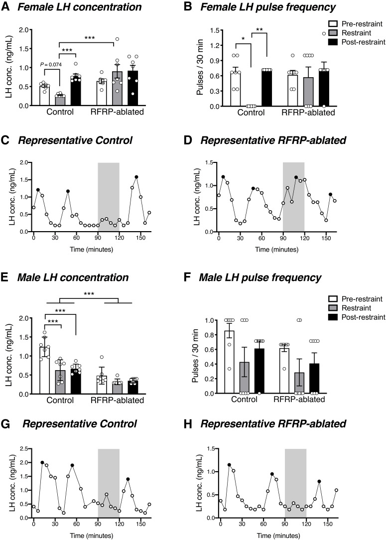
Figure 6.
RFRP neuronal ablation prevents acute stress-induced LH suppression. A, B, In response to a 30 min restraint stress, female control mice, but not RFRP-ablated mice, exhibited a strong trend toward stress-induced suppression in average circulating LH concentration (p = 0.074 vs the 90 min pre-restraint period, and p = 0.001 vs the 45 min post-restraint period) and a significant suppression in LH pulse frequency (p = 0.048 vs the pre-restraint period, and 0.004 vs the post-restraint period) (n = 7 per group). During restraint, LH concentration was significantly lower in control females than in RFRP-ablated mice (p = 0.0002). C, D, Representative examples of LH profiles in a control and RFRP-ablated female mouse. Gray shaded bars represent the 30 min restraint stress. E, Control male mice exhibited a significant stress-induced suppression in basal LH (p < 0.0001) that was not observed in RFRP-ablated mice (p = 0.072), yet (F) neither control nor RFRP-ablated male mice showed a stress-induced change in LH pulse frequency (n = 7 per group). G, H, Representative examples of LH profiles in a control and RFRP-ablated male mouse. Two-way ANOVA and Holm-Sidak's multiple comparisons tests were used for analysis of LH concentration, while effects of restraint stress on LH pulse frequency were analyzed using the Friedman test followed by Dunn's multiple comparison test. Data are mean ± SEM. Black circles represent pulse peaks.

