
Fig. 6. Requirement of H3.3A for neural crest–derived glia and pigment cells.
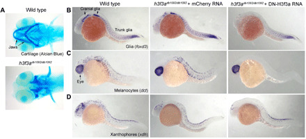
(A) Ventral whole-mount views of larval zebrafish heads at 5 dpf stained with Alcian Blue. Homozygous h3f3adb1092 mutants display complete loss of neural crest–derived jaw cartilages (n = 10/10). (B to D) In situ hybridization of zebrafish embryos for markers of glia (foxd3; 24 hpf), melanocytes (dct; 27 hpf), and xanthophores (xdh; 27 hpf). Homozygous h3f3adb1092 mutants injected at the one-cell stage with a control mCherry RNA show partial reductions in cranial glia (n = 5), melanocytes (n = 4), and xanthophores (n = 3), while those injected with dominant-negative H3f3a RNA to further reduced H3.3A function show complete loss of melanocytes (n = 5) and severe reductions of glia (n = 6) and xanthophores (n = 4) throughout cranial and trunk regions.