
Fig. 3. Dabrafenib mitigates cisplatin-activated BRAF signaling cascade.
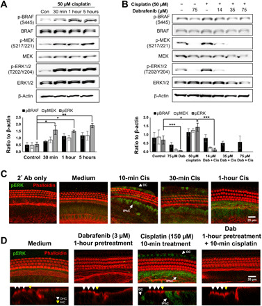
(A) Representative Western blot images of BRAF, ERK, and MEK phosphorylation in HEI-OC1 cells upon 50 μM cisplatin treatment for 30 min, 1 hour, and 5 hours. Phosphorylated protein bands were normalized to β-actin and averaged, means ± SEM, *P < 0.05, **P < 0.01 by one-way ANOVA with Bonferroni post hoc test. n = 4. (B) Representative Western blot images (n = 3) of BRAF, ERK, and MEK phosphorylation upon combined dabrafenib (14, 35, or 75 μM) and cisplatin (50 μM) treatment in HEI-OC1 cells. Cells are pretreated with dabrafenib for 1 hour before 1-hour cisplatin treatment. Medium alone, cisplatin alone, and 75 μM dabrafenib alone used as controls. Phosphorylated protein bands were normalized to β-actin and averaged, means ± SEM, *P < 0.05, ***P < 0.001 by one-way ANOVA with Bonferroni post hoc test. n = 3. (C) Representative phalloidin (red) and phosphorylated ERK (pERK) (green) stained confocal images of P3 FVB whole-mount middle turn mouse cochlea explants pretreated with 3 μM dabrafenib (Dab) for 1 hour before 10 min cisplatin (150 μM) exposure. Deiters’ cells (DC) and inner phalangeal cells (IPhC) with labeled arrows. n = 6 cochlea. (D) Representative phalloidin (red)– and pERK (green)–stained confocal images of P3 FVB whole-mount middle turn mouse cochlea explants pretreated with 3 μM dabrafenib (Dab) for 1 hour before 10 min cisplatin (150 μM) exposure. Ortho section shown below in which OHCs are identified with white arrows, inner HCs (IHCs) are identified with yellow arrows, and pERK-positive DCs and IPhCs are identified with labeled arrows. n = 6 cochlea.