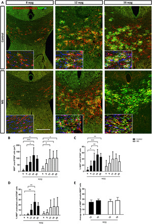
Fig. 3. MS-hiOL–derived progeny extensively myelinates the dysmyelinated Shi/Shi:Rag2−/− corpus callosum.

(A) Combined detection of human nuclei (STEM101) and human cytoplasm (STEM 121) (red) with MBP (green) in the Shi/Shi Rag2−/− corpus callosum at 8, 12, and 16 wpg. General views of horizontal sections at the level of the corpus callosum showing the progressive increase of donor-derived myelin for control- (top) and MS- (bottom) hiOLs. (B) Evaluation of the MBP+ area over STEM+ cells. (C and D) Quantification of the percentage of (C) MBP+ cells and (D) MBP+ ensheathed cells. (E) Evaluation of the average sheath length (μm) per MBP+ cells. No obvious difference was observed between MS and control-hiOLs. Two-way ANOVA followed by Tukey’s multiple comparison tests were used for the statistical analysis of these experiments (n = 6 to 14 mice per group). Error bars represent SEMs. *P < 0.05, **P < 0.01, and ***P < 0.001. Scale bar, 200 μm. See also figs. S3 and S5.
