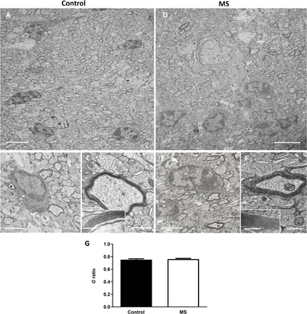
Fig. 4. MS-hiOLs produce compact myelin in the dysmyelinated Shi/Shi:Rag2 −/− corpus callosum.

(A to F) Ultrastructure of myelin in sagittal sections of the core of the corpus callosum 16 wpg with control-hiOLs (A to C) and MS-hiOLs (D to F). (A and D) General views illustrating the presence of some electron dense myelin, which could be donor derived. (B, C, E, and F) Higher magnifications of control (B and C) and MS (E and F) grafted corpus callosum validate that host axons are surrounded by thick and compact donor derived myelin. Insets in (C) and (F) are enlargements of myelin and show the presence of the major dense line. No difference in compaction and structure is observed between the MS and control myelin. (G) Quantification of g-ratio revealed no significant difference between myelin thickness of axons myelinated by control- and MS-hiOLs. Mann-Whitney t tests were used for the statistical analysis of this experiment (n = 4 mice per group). Error bars represent SEMs. Scale bars, (A and D) 5 μm , (B and E) 2 μm, and (C and F) 500 nm [with 200 and 100 nm, respectively in (C) and (F) insets].
