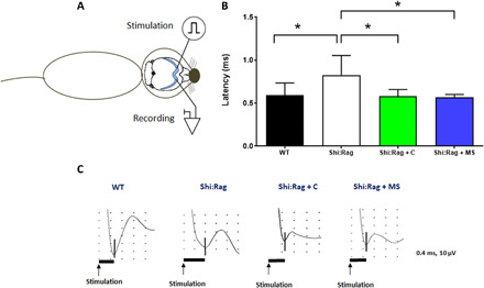
Fig. 5. MS-hiOLs improve transcallosal conduction of the dysfunctional Shi/Shi:Rag2−/− axons to the same extent than control-hiOLs.

(A) Scheme illustrating that intracallosal stimulation and recording are performed in the ipsi- and contralateral hemisphere, respectively. (B) N1 latency was measured following stimulation in different groups of Shi/Shi:Rag2−/−: intact or grafted with control or MS-hiOLs and WT mice at 16 wpg. MS-hiOL–derived myelin significantly restored transcallosal conduction latency in Shi/Shi:Rag2−/− mice to the same extent than control-derived myelin (P = 0.01) and close to that of WT levels. One-way ANOVA with Dunnett’s multiple comparison test for each group against the group of intact Shi/Shi:Rag2−/− was used. Error bars represent SEMs. *P < 0.05. (C) Representative response profiles for each group. Scales in Y axis is equal to 10 μV and in the X axis is 0.4 ms.
