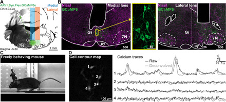
Fig. 1. Activity of V2a neurons can be recorded with micro-endoscopy.

(A) Schematic of viral injections and lens implantation into the Gi of a Chx10-Cre animal for calcium imaging in freely behaving mice. (B) Nissl-stained (magenta) coronal sections of Chx10-Cre mouse showing native GCaMP6s fluorescence (green) and the location of medial (left) or lateral (right) GRIN lens implants. Inset in the yellow box is a magnified view of GCaMP6s expression in V2a neurons. Scale bars in micrometers. (C) Example mouse during treadmill running with miniature microscope attached. Photo credit: Joanna Schwenkgrub, CNRS. (D) Example cell contour map (left) with corresponding raw/deconvolved calcium traces (right). Abbreviations used in (A) and (B): 4V, 4th ventricle; 7N, facial motor nucleus; 7n, facial nerve; py, pyramidal tract; Gi, gigantocellular reticular nucleus; sp5, spinal trigeminal tract; a.u., arbitrary units.
