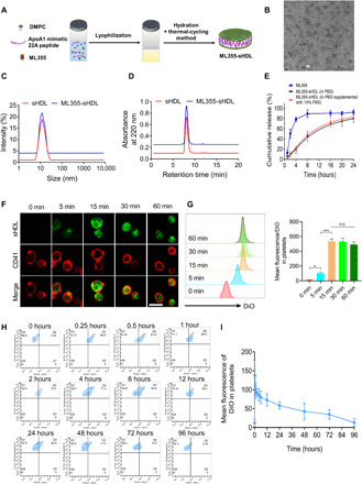
Fig. 1. Preparation and characterization of ML355-sHDL.

(A) Illustration of the synthesis of ML355-sHDL composed of an ApoA1 mimetic 22-mer peptide (22A), 1,2-dimyristoyl-sn-glycero-3-phosphocholine (DMPC), and ML355 using the thermal-cycling method. (B) Negative-stained transmission electron microscopy image of ML355-sHDL; scale bar, 20 nm. (C) Dynamic light scattering of blank sHDL and ML355-sHDL. (D) Gel permeation chromatography of blank sHDL and ML355-sHDL. (E) Release of ML355 from sHDL at PBS and PBS cumulative supplemented with 10% fetal bovine serum (FBS). Data are means ± SD (n = 3). (F) Platelet uptake and intracellular distribution of DiO-sHDL in platelets imaged by confocal microscopy (scale bar, 2 μm). Washed mouse platelets were suspended in 1 ml of Tyrode’s buffer at 3 × 10⁶ platelets/ml and incubated with DiO-sHDL (sHDL at 50 μg/ml and DiO at 2.5 μg/ml) for indicated lengths of time (5, 15, 30, and 60 min) at 37°C. Platelets were then washed with Tyrode’s buffer twice to remove free DiO-sHDL and then fixed with 4% paraformaldehyde, followed by staining with Alexa Fluor 647–conjugated anti-mouse CD41 antibody. Platelet membrane is shown in red, and sHDL particle accumulation in platelets is shown in green. (G) Representative flow cytometry histograms and data analysis of mean DiO fluorescence intensity in platelets by flow cytometry. Data are means ± SD (n = 3). *P < 0.05 and N.S. (no significant difference). (H) CD41-PE–positive platelets in the blood versus DiO-sHDL. (I) Quantification of DiO-sHDL uptake by CD41-PE–positive platelets in blood. Data are means ± SD (n = 4).
