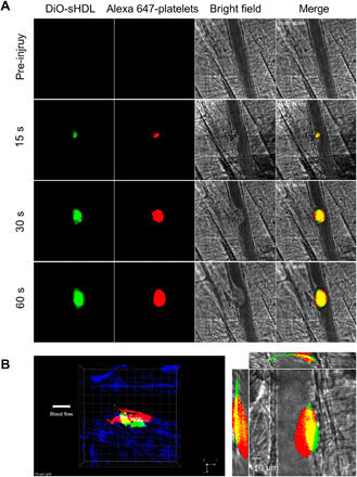
Fig. 3. sHDL targets thrombus in vivo.

Male mice were pretreated with DiO-sHDL via intravenous injection with sHDL (50 mg/kg) and DiO (2.5 mg/kg). After 24 hours, mice were anesthetized and surgically prepared as described in detail in Materials and Methods. DyLight 647–conjugated rat anti-mouse platelet GP1bβ antibody (0.1 μg/g, X649, EMFRET Analytics) was administered by jugular vein cannula before vascular injury. Multiple independent thrombi (8 to 10) were induced in the arterioles (30 to 50 μm diameter) per mouse (three mice in each group) by a laser ablation system as described in Materials and Methods. Images of thrombus formation at the site of injured arterioles were acquired in real time under 63× water-immersion objective with a Zeiss Axio Examiner Z1 fluorescence microscope. (A) Sequence of intravital fluorescence microscopic images recorded over 1 min showing accumulation of fluorescently labeled DiO-sHDL in a forming thrombus indicated by fluorescently labeled platelets. (B) Left: Representative three-dimensional confocal images of DiO-sHDL (green) and platelet accumulation (red) in stable thrombi recorded under confocal intravital microscopy. Right: Representative middle section of thrombus shown under a three-direction view.
