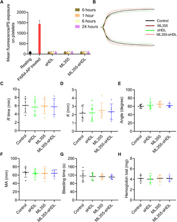
Fig. 6. ML355-sHDL does not affect the phosphatidylserine exposure on platelet surface, coagulation system, and hemostatic action.

Both female and male mice (n = 6) were pretreated intravenously with saline control, sHDL (50 mg/kg), ML355 (1.5 mg/kg), or ML355-sHDL (sHDL at 50 mg/kg and ML355 at 1.5 mg/kg). All treated groups were subjected to the following different examinations. (A) At different time points after administration (1, 6, and 24 hours), the phosphatidylserine (PS) exposure over the time course in platelets from different groups was quantified by flow cytometry. Blood collected from untreated mice was taken as resting platelets (negative control), and blood stimulated with protease-activated receptor 4–activating peptide (PAR4-AP) (200 μM) was used as activated platelets (positive control). (B) Representative tracings indicating different treatments were presented. Some major coagulation parameters were analyzed. (C) R time, time to formation of the initial fibrin threads (min). (D) K time, the time until the clot reaches a certain strength (min). (E) Alpha (α) angle, the rapidity with which the clot forms (degree). (F) Maximum amplitude (MA), clot’s maximum strength (mm). (G) Bleeding times and (H) blood loss (quantified as milligrams of hemoglobin) were quantified.
