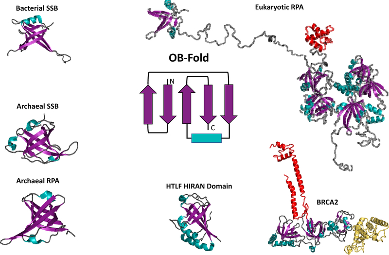
Figure 2: Oligonucleotide/oligosaccharides binding folds (OB-folds) are conserved across all domains of life.
The cartoon at the center of the figure is the basic OB-fold motif. While there is some variation, the five beta sheets (purple) that form the mixed beta barrel and the alpha helix (teal) that caps the barrel are generally conserved. OB-folds are represented in each of the structures (bacterial SSB, archaeal SSB, archaeal RPA, and eukaryotic RPA and are also present in numerous ssDNA binding proteins represented here by the Hiran domain of HLTF and the ssDNA binding domain of human BRCA2) using the same purple beta sheet/teal alpha helix/grey loops scheme. Additional structural components are colored differently to highlight them. In the eukaryotic RPA structure the winged helix (WH) domain is depicted in red. In the BRCA2 structure the tower domain is depicted in red and the helix domain is depicted in yellow.

