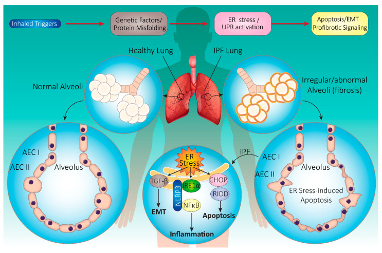
Figure 3.
Schematic summary of IPF pathogenesis. Inhaled triggers along with genetic factors contribute to the induction of UPR in IPF pathogenesis. UPR activation triggers many downstream pathways, which may induce EMT, apoptosis, and pro-fibrotic signaling in the lung. The key histopathological feature of IPF is usually interstitial pneumonia (UIP). Endoplasmic reticulum (ER) stress in IPF triggers apoptosis through UPR system activation, inflammation via nuclear factor kappa-light-chain-enhancer of activated B cells (NFκB) activation, and epithelial–mesenchymal transition (EMT) through transforming growth factor-β (TGF-β) activation. Affected cells include alveolar epithelial cell (AEC) I and II, resident macrophages, and fibroblasts in the alveoli. Alveolar changes lead to exaggerated fibrosis, lymphocyte infiltration, and honeycomb change. Abbreviations: CHOP (C/EBP-homologous protein), IRE1 (serine/threonine-protein kinase/endoribonuclease inositol-requiring enzyme 1), RIDD (regulated IRE1-dependent decay), NLRP3 (NOD-, LRR-, and pyrin domain-containing protein 3).

