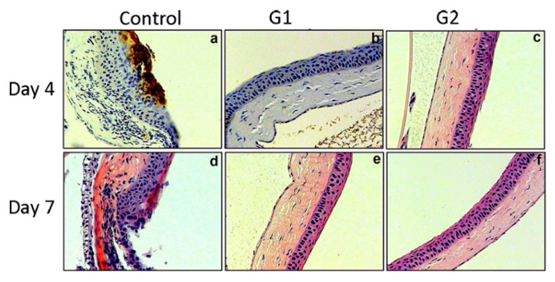
Figure 4.
A mouse model of corneal keratitis was given 100 μL of 0.5mM of G1 (peptides with alternating charges), G2 (peptides with repetitive charges), and designated peptide (control) as a prophylactic eye drop followed by the inoculation of HSV-1. Immunohistochemistry was carried out using anti-HSV-1 glycoprotein D (gD) polyclonal antibody on day 4 and day 7 post-infection to detect the HSV-1 gD expression in the cornea. (a) chronic inflammation was observed together with significant brown staining in the pretreated cornea with control on day 4, which was indicating the expression of HSV-1 gD; (b) on day 4, HSV-1 gD staining was not detected in the cornea pretreated with G1 peptide; (c) on day 4, HSV-1 gD staining was not detected in the cornea pretreated with G2 peptide; (d) the staining was disappeared in the cornea pretreated with control on day 7 but the damage of the corneal epithelium was observed; (e) on day 7, HSV-1 gD staining was not detected in the cornea pretreated with G1 peptide and the corneal epithelium remained intact; (f) on day 7, HSV-1 gD staining was not detected in the cornea pretreated with G2 peptide and the corneal epithelium remained intact. The results indicated G1 and G2 significantly blocked the entry of HSV-1, adapted with permission from [85], American society for biochemistry and molecular biology, 2011.

