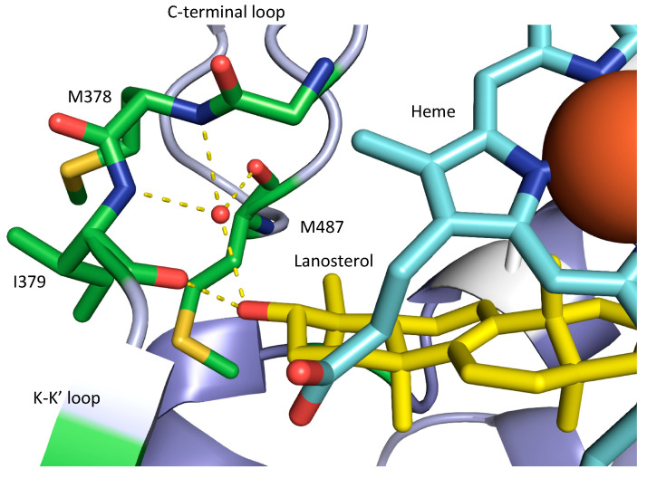
Figure 2.
Structural view of the ligand-binding pocket of the human CYP51 D231A H314 mutant catalytic domain (Protein Data Bank ID: 6UEZ) interacting via hydrogen bonds (yellow dashes) with the hydroxyl of lanosterol (carbons in yellow). The heme is given in blue with the iron as a large red ball. Amino acid residues involved in the hydrogen bonding directly or as part of a water-mediated network are indicated. A list of all key AA residues for the main structures of the SDM are given in Supplementary Table S3.

