
Figure 2.
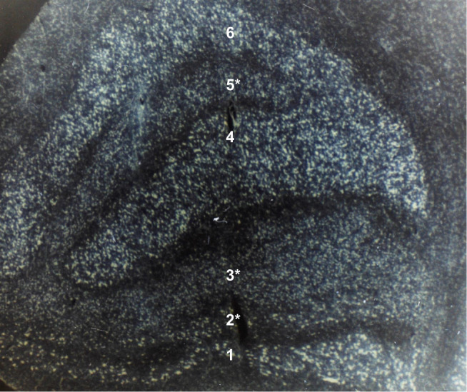
Coronal section of the left lateral geniculate nucleus in a 72-year-old patient after 8 years of optic atrophy from absolute glaucoma in the left eye. The smaller cells with less staining in ipsilateral layers 2, 3, and 5 (marked with an asterisk) represent transsynaptic atrophy. Layers 1 and 2 are the magnocellular layers. Layers 3 to 6 are the parvocellular layers. (Contrast-enhanced grayscale photo of a Nissl-stained specimen at 20x magnification; unpublished observation).