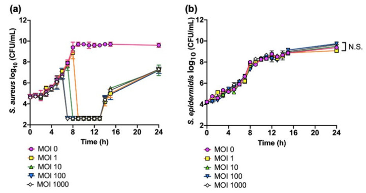
Figure 1.
Killing assays of S. aureus SA-1 and S. epidermidis SE-4 treated with phage SaGU1. (a) S. aureus SA-1 lysis with phage SaGU1 infection. (b) S. epidermidis SE-4 growth in the presence of phage SaGU1. The culture was started with ~104 CFU/mL of staphylococci and appropriate amounts of phage SaGU1. The data are presented as the mean ± SD of at least three independent experiments. The detection limit was 4.0 × 10² CFU/mL. Student’s t-test was used to determine a significant difference between the presence and absence of phage SaGU1. N.S.; not significant.

