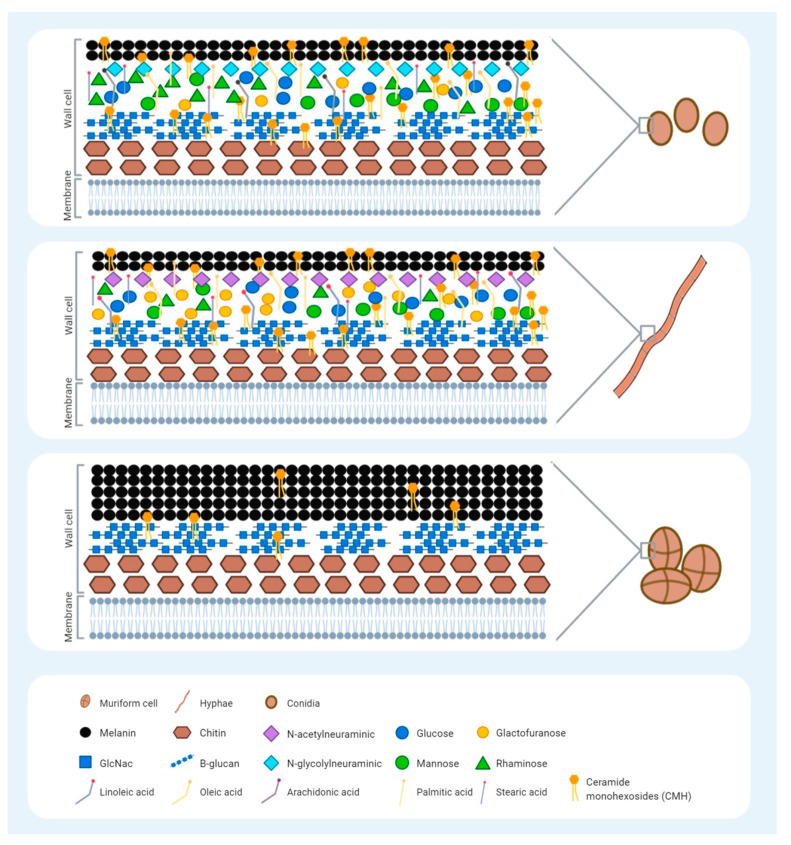
Figure 1.
Schematic representation of the Fonsecaea pedrosoi cell wall, highlighting the reported molecules in each morphological form: conidia, hyphae, and muriform cells. The fungal cell wall is a heterogenic structure that is important for activation of the host innate immune system during infection through the pathogen-associated molecular patterns (PAMPs) recognized by the Pattern-recognition receptors (PRRs) present on innate immune cells. The morphological forms of F. pedrosoi have similarities but also differences with regard to the type and amount of the molecules present. No difference in chitin and B-glucan expression have been reported for the three different forms of F. pedrosoi. Conidia predominantly express sialic acid N-glycolylneuraminicacid, whereas hyphae express N-acetylneuraminicacid. Carbohydrates, such as glucose, mannose, and glucosamine, are similarly deposited on conidia and hyphae surfaces; however, conidia have greater expression of rhamnosis, while hyphae have greater galactofuranosis expression. Conidia also present a greater amount of lipids than hyphae, and the expression of the arachidonic fatty acids seems to be exclusive to the conidia form. The expression of linoleic fatty acids is exclusive to the hyphae, while the palmitic, stearic, and oleic fatty acids are present in both morphologies. Ceramide monohexosides (CHM) consist of a carbohydrate unit bound to a ceramide with antigenic properties. Presenting on the surface of all three F. pedrosoi morphologies, it is related to fungal differentiation. Melanin is a pigment present in the outer layer of all forms of F. pedrosoi, which provides their typical dark brown or black stain. A higher deposition of melanin is observed in muriform cells that block the binding sites of the anti-CHM antibodies, contributing to a greater virulence and immune evasion in these cells.

