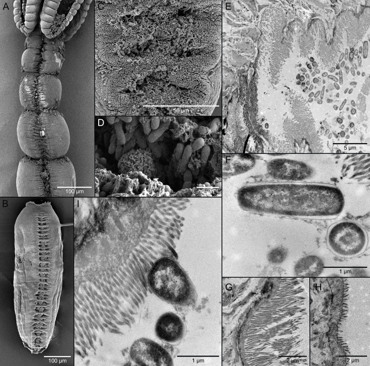
Fig 2. Scanning (SEM) and transmission (TEM) electron micrographs of Caulobothrium multispelaeum (Cestoda: Tetraphyllidea).
(A) SEM of anterior portion of worm showing grooves of symbiotic organ in immature proglottids. (B) SEM of mature proglottid showing tandem series of elliptical apertures of symbiotic organ; white rectangle indicates location of detail in C. (C) SEM of apertures of symbiotic organ. (D) SEM of bacillus-like bacteria in apertures of symbiotic organ. (E) TEM of cross section through portion of symbiotic organ showing bacteria in cavity. (F) TEM of bacteria in cavity in frontal and cross section. (G) TEM of elongate filitriches lining surface of cavity of symbiotic organ. (H) TEM of shorter filitriches on other body surfaces. (I) TEM of bacterium in intimate contact with filitriches lining cavity of symbiotic organ.

