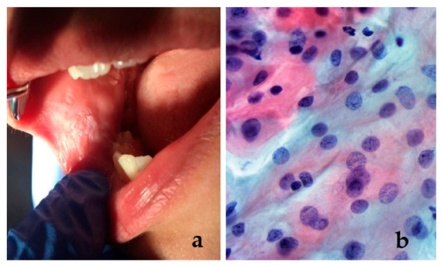
Figure 3.

(a) A 12-year-old female patient with a white “patch” on the buccal mucosa, resembling leukoplakia; histopathological examination was performed in order to obtain the correct diagnosis; (b) exfoliative cytology did not confirm the presence of a premalignant lesion. Intermediate squamous cells (reflecting the accelerate turnover) with slight inflammation (different shape of nuclei, stainability, irregular contour of the nuclear border) (Papanicolaou stain, ×40).
