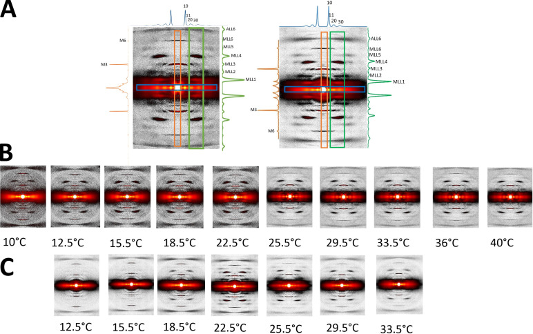
Figure 2.
XRD patterns from tarantula skeletal muscle. (A) Relaxed muscles of an ex vivo leg (left) and an isolated live muscle (right), recorded at 22.5°C. Equatorial, meridional, and off-meridional intensity profiles (as shown in the corresponding colored boxes) are seen in blue (top), orange (left), and green (right), respectively. The axial span of the blue box in the equator is ±0.00755 nm−1; the radial span of the orange box in the meridian is ±0.0081 nm−1; and the green box radial range span is from 0.038 nm−1 to 0.072 nm−1. (B and C) XRD patterns of relaxed ex vivo leg muscles (B) and isolated muscles (C) recorded from 10°C to 40°C.

