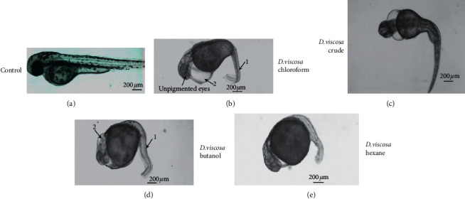
Figure 5.

D. viscosa induced severe teratogenicity in zebrafish embryos. Representative micrograph of live zebrafish embryos mock treated (control) and treated with crude extract and various fractions of D. viscosa. The images were recorded after two days of exposure to zebrafish embryos to sub-lethal concentration of crude and various fractions. The images are arranged in order from mild to severe abnormalities, being the crude extract. The presence of pectoral fin mock treated embryos shows that the embryos are not developmentally delayed. The mock treated embryos have straight body with straight arrangement of notochord (black arrow, notochord in control) and pigments all around the body and also have pigment development in eyes, whereas the treated embryos lack the pigmentation and their body is curved due to abnormal development of notochord (represented by “1” in treated embryos). The treated embryos have large cardiac edema (2 in all the treated embryos). The pigment is also lacking in treated embryos except the embryos which were treated with crude extract.
