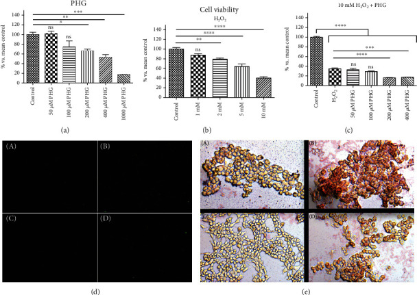
Figure 1.

The effect of PHG on cell viability and steatosis. (a, c) PHG was nontoxic, both referring to the control and H2O2 in concentrations below 200 μM and dose-dependently lowered cell viability in concentrations above 200 μM. (b) H2O2 showed a dose-dependent effect on cell viability with GI50 around 10 mM. (d) Immunofluorescence staining of active Caspase 3 (A: control group; B: NAFLD; C: PHG; D: NAFLD+PHG). (e) Oil Red O staining of HepG2 cells (A: control group; B: NAFLD; C: PHG; D: NAFLD+PHG).
