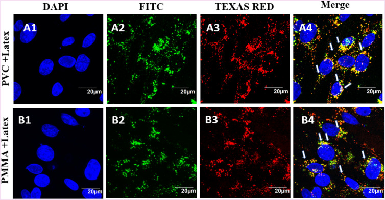
Figure 6.
BHK-21 cells exposed to PVC (A1–A4) and PMMA (B1–B4) NPs at a concentration of 200 µg/mL for 24 h and latex beads (80 µg/mL). The images represent nucleus stained with Hoechst dye and cells exposed to PVC or PMMA NPs (green emitting, A2, B2) and red fluorescent latex beads (A3, B3). The blue, green and red fluorescent images are overlaid to show the localization of PVC and PMMA NPs and latex beads inside the cell, indicating that PVC and PMMA NPs are entering the cell by endocytosis.

