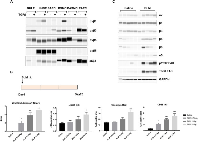
Figure 1.
Changes of αv integrin expression upon fibrosis induction in the lung. (A) The expression of αv integrins in various human primary lung cell types upon TGFβ (5 ng/ml for 24 h) treatment. Following immunoprecipitation with an anti-αv antibody, the αvβ1, αvβ3, αvβ5, and αvβ6 heterodimers were detected by Sally Sue simple western analysis after using antibodies that recognize each individual β-subunit. Normal human lung fibroblast, NHLF; normal human bronchial epithelial cells, NHBE; small airway epithelial cells, SAEC; bronchial smooth muscle cells, BSMC; pulmonary artery smooth muscle cells, PASMC; pulmonary artery endothelial cells, PAEC. Full-length blot images are presented in Supplemental Fig. S6A–E. (B) Development of a bleomycin-induced lung fibrosis model in mice. Bleomycin (BLM) was administered at the indicated doses via intra-tracheal (i.t.) instillation. After 20 days, lungs were collected for histological analyses. Modified Ashcroft score, Picosirus red staining, immunohistochemical analyses of αSMA and CD68 of total lung were quantified and shown (mean ± SEM, n = 5). Immunohistochemistry, IHC. One-way ANOVA followed by Tukey’s test, *p < 0.05, **p < 0.01, ***p < 0.005 vs Saline group. (C) Integrin expression and signaling in fibrotic lungs (BLM 0.5U/kg bw) was determined by Sally Sue simple western analysis using antibodies that recognized the individual α or β-subunits. Each lane represents total lung homegenate for one animal, n = 5 for saline or BLM group. GAPDH level in total lung lysates was used as a loading control. Full-length blot images are presented in Supplemental Fig. S6F–N.

