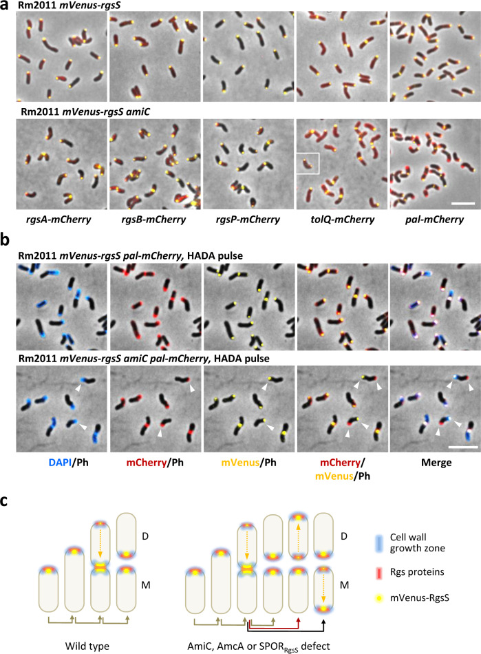
Fig. 3. Rgs proteins, TolQ and PG incorporation zones colocalize with RgsS in both wild type and amiC cells.
a Fluorescence microscopy images of exponentially growing Rm2011 mVenus-rgsS and Rm2011 mVenus-rgsS amiC cells, producing mCherry fusions of the indicated proteins from gene fusions at the native genome locations. Cell samples were taken from exponential phase TY cultures. The insert shows an additional cell of the same strain representative of cells with bipolar colocalization of mVenus-RgsS and TolQ-mCherry fluorescence foci. The images are representative of two independent cultivations and microscopy analyses. Scale bar, 5 µm; Ph phase contrast. b Fluorescence microscopy of Rm2011 mVenus-rgsS and Rm2011 mVenus-rgsS amiC cells, carrying pal-mCherry at the native genomic location, pulse-labeled with HADA for 3 min. Cell samples were taken from exponential phase TY cultures. Arrowheads show cells with non-colocalized mVenus-RgsS and Pal-mCherry. Scale bar, 5 µm; Ph phase contrast. The images are representative of three independent cultivations, HADA staining and microscopy analyses. c Schematic representation of cell growth polarity inheritance inferred from data shown in this figure, Fig. 2 and Supplementary Figs. 10–12 and 18. In wild-type cells, Rgs proteins and zones of PG biosynthesis are inherited to the new cell pole. In cells, lacking AmiC, AmcA, or the SPOR domain of RgsS, the Rgs proteins and PG synthesis zones are occasionally observed at the old cell pole, representing cells with inverted growth polarity. D daughter cell, M mother cell.

