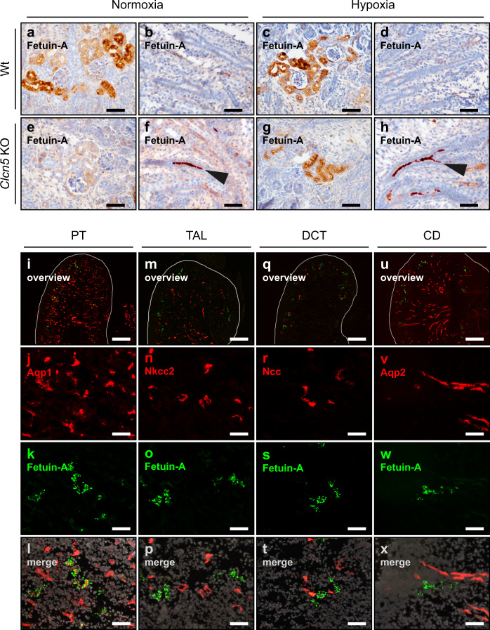
Fig. 3. Fetal hypoxia induces fetuin-A expression in the proximal tubulus.
a–h Fetuin-A staining on E18.5 kidney sections of normoxic (a, b, e, f) or hypoxic (c, d, g, h), wild type (a–d) or Clcn5 KO (e–h) fetuses. Arrowheads indicate intraluminal fetuin-A staining that results from the impaired endocytosis of low molecular weight proteins in the PT of Clcn5 KO mice (f, h). i–x Immunofluorescence staining of the indicated nephron segment marker proteins (red) and fetuin-A (green) on E18.5 kidney sections. PT, proximal tubulus (i–l); TAL, thick ascending limb (m–p); DCT, distal convoluted tubulus (q–t); CD, collecting duct (u–x). Images are representative of at least three independent antibody stainings (a–x). Scale bar = 50 µm (a–x), except overview images for which the scale bar = 300 µm.

