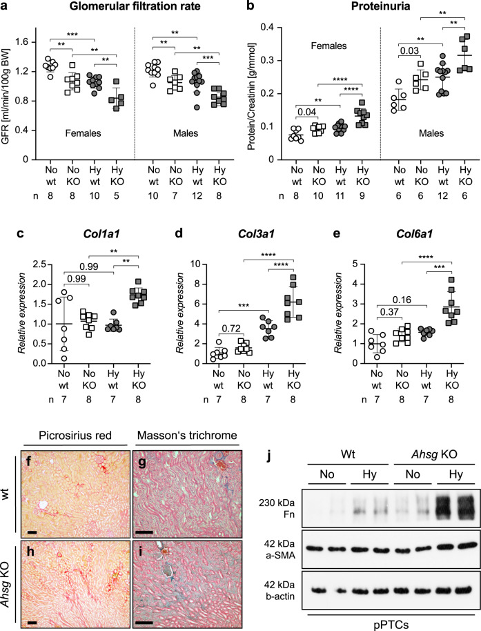
Fig. 5. Fetuin-A deficiency aggravates CKD progression in hypoxic IUGR kidneys.
a, b Decline of renal function in adult hypoxic offspring showed additive effects of hypoxia and fetuin-A deficiency. The decline in GFR was indistinguishable between the sexes with the greatest functional reduction in hypoxic Ahsg KO animals (a). The incline in proteinuria (protein/creatinine ratio) was more pronounced in males than in females. For both sexes, hypoxic Ahsg KO animals had the highest ratios (b). Male and female samples are analyzed separately. c–e Relative mRNA expression levels of Col1a1 (c), Col3a1 (d), and Col6a1 (e) were markedly enhanced in kidneys of hypoxic Ahsg KO offspring. f–i Histological depiction of collagen using picrosirius red (f, h) or Masson’s trichrome staining (g, i) showed a stronger, more intricate pattern on kidney sections of hypoxic Ahsg KO offprings compared to controls. Images are representative of at least three independent experiments. Scale bar = 100 µm. j Primary proximal tubular cells (pPTCs) isolated from two different wt or Ahsg KO mice exhibit enhanced expression of fibronectin and α-smooth muscle actin (α-SMA) protein upon culture in hypoxic conditions. Images are representative of three independent Western blots. Uncropped blots in Source Data. Data were analyzed from N = hypoxic or normoxic offspring and are presented as mean ± SEM (a–e). Ordinary one-way ANOVA with Dunnett’s multiple comparisons test (a–b) or Ordinary one-way ANOVA with Tukey’s multiple comparisons test (c–e). Individual P-values are denoted above the comparison lines (b, c, e). (****P < 0.0001; ***P < 0.001; **P < 0.01). Source data are provided as a Source Data file.

