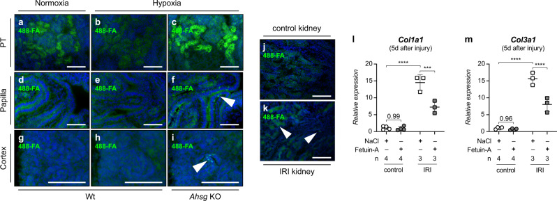
Fig. 8. Fetuin-A supplementation reduces the expression of fibrotic markers upon hypoxia-related injury.
a–i Fetuin-A deficiency promotes accumulation of calcium mineral particles in hypoxic fetal kidneys. Calcium biominerals were detected by ATTO 488 fluorescently labeled fetuin-A (488-FA). Compared to normoxic or hypoxic wt mice, hypoxic Ahsg KO mice exhibited the strongest 488-FA staining intensity in the proximal tubulus (PT), indicative of an increased mineralized matrix turnover (a–c). Arrowheads in f and i point towards granular staining pattern in the papilla and cortex, respectively, reflecting bulk accumulation of 488-FA in kidneys of hypoxic Ahsg KOs. Such granules were not detectable in wt samples (d, e, g, h). j, k Ischemia-reperfusion injury (IRI) induces calcium mineral particles in adult kidneys. A granular staining pattern indicative of bulk accumulation of 488-FA at sites of calcium deposits was only present in IRI kidneys (k), but not in controls (j). l, m Fetuin-A supplementation reduced the expression of the fibrotic markers Col1a1 (l) and Col3a1 (m) in IRI kidneys 5 days after injury. No effect is seen in mice treated with physiological saline solution (NaCl). Data were analyzed from N = kidneys and are presented as mean ± SEM. Ordinary one-way ANOVA with Dunnett’s multiple comparisons test. Individual P-values are denoted above the comparison lines. (****P < 0.0001; ***P < 0.001). Images are representative of at least three independent experiments (a–k). Scale bar = 100 µm (a–k). Source data are provided as a Source Data file.

