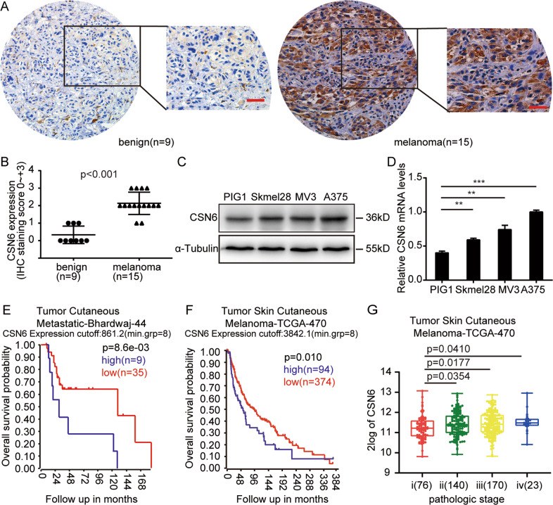
Fig. 1. High CSN6 expression is associated with a poor melanoma patient prognosis.
A Representative immunohistochemical staining assays showing CSN6 expression in human benign nevus tissue (left, n = 9) and melanoma tissue (right, n = 15). B Immunohistochemical analyses of CSN6 expression levels in 9 benign nevus tissue samples and 15 melanoma tissue samples, P < 0.001. C, D Western blot and qRT-PCR assays performed to detect CSN6 expression in the A375, MV3, and Skmel28 melanoma cell lines and the immortalized melanocyte cell line PIG1. E, F Kaplan–Meier analysis of the overall survival probability using data from the online R2 database. P-values calculated by the log-rank test are indicated. G Box plot analysis of CSN6 expression levels with the pathological stage of melanoma.

